
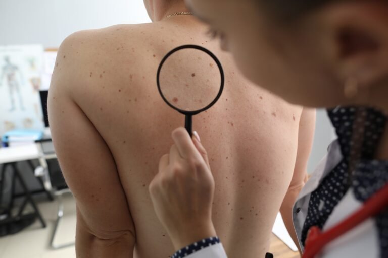
Skin cancer develops when skin cells undergo abnormal and rapid multiplication, typically in the layer of the skin known as the epidermis. Identifying these cellular changes promptly can lead to more effective management and care. Understanding the basic mechanisms behind this disease helps individuals remain vigilant about their health. The condition affects people across all demographics, and regular observation of your skin provides a straightforward method for detecting early alterations.
What Is Skin Cancer?
The human body constantly produces new skin cells to replace older ones. When DNA mutations occur within the epidermis, this structured process fails, leading to unchecked cellular division. This rapid multiplication forms a mass of abnormal cells called a tumor, and is the physical manifestation of skin cancer. Ultraviolet light exposure from the sun acts as the most frequent cause for these DNA changes. Frequent sunburns and the use of indoor tanning beds increase the risk of developing the disease. Other risk factors include having a fair complexion, naturally light hair, blue or green eyes, or a family history of the condition.
Medical professionals generally categorize the disease into a few main types. Basal cell carcinoma typically appears on sun-exposed areas, like the face and neck. Squamous cell carcinoma also surfaces on frequently exposed skin, but it can present differently. Melanoma originates in the pigment-producing cells and is a very aggressive form of the disease.
What Are the Signs?
Detecting abnormalities early requires knowing what specific changes to look for. Each major type of the disease presents with distinct visual characteristics. Basal cell carcinoma may resemble a small, slightly transparent, or pink bump on the skin’s surface; it grows slowly over time. While it rarely spreads to distant organs, it can cause significant damage to the surrounding healthy tissue if left unaddressed.
Squamous cell carcinoma often materializes as a firm, red nodule or a flat lesion, featuring a scaly, crusted surface. Patients might notice these sores developing on the ears, face, back, or neck. This form can be more aggressive than basal cell carcinoma and possesses the capacity to spread to other parts of the body.
Melanoma usually begins as a new mole or presents as a noticeable change in an existing one. The indicators for melanoma may include asymmetrical shapes, irregular borders, shifting colors, and a larger-than-normal diameter. Because melanoma can spread rapidly through the body, early detection remains a primary focus of dermatological screenings.
How Is It Treated?
Dermatologists determine the most appropriate intervention based on the specific type. Surgical excision involves cutting out the cancerous tissue along with a margin of healthy skin to clear the area of disease. For more complex or recurrent cases, physicians might utilize Mohs surgery. This precise technique removes the visible tumor and sequentially targets microscopic layers of skin, examining each layer until no cancer cells remain. Cryotherapy utilizes liquid nitrogen to freeze and eliminate small, early-stage growths. Photodynamic therapy combines specialized light-sensitive topical medications with a specific light source to target and eliminate the diseased tissue.
Seek Professional Dermatology Care
Proactive monitoring of your skin serves as a primary defense against advancing disease, allowing you to detect changes early. If you observe a suspicious growth, a sore that fails to heal, or a mole that changes appearance, schedule an evaluation with a dermatologist. Professionals possess the specialized training needed to accurately diagnose skin conditions and create a tailored treatment strategy. Early medical intervention consistently yields the most favorable outcomes for patients.